Some cavities show obvious signs: a dark spot, a small hole, sensitivity to cold or pain. Others, however, can develop over a long period without giving any indication when you look in the mirror.
So-called proximal or interproximal cavities occur on the surfaces where two teeth come into contact. This is precisely one of the most difficult areas to see and also one of the most difficult to clean using a toothbrush alone.
This helps to explain a situation that surprises many patients: during a check-up, the teeth appear normal at first glance, but an X-ray reveals a lesion between two of them.
It doesn’t mean that the cavities appeared out of the blue. It simply means that they were developing in a place that’s hard to see.
What happens in that tiny space between two teeth?
Cavities don’t start out as holes. It’s a process.
A biofilm consisting of bacteria, components of saliva and other elements continuously forms on the surface of the teeth. When these bacteria metabolise certain sugars from food, they produce acids that temporarily lower the pH level near the enamel.
The enamel then loses minerals. Saliva and fluoride can help restore some of these minerals between episodes, which is why there is a constant balance between demineralisation and remineralisation.
The problem arises when this balance remains tipped towards mineral loss for too long.
There is an additional challenge between two teeth: a conventional toothbrush cannot effectively clean the surface immediately below the point of contact. If biofilm frequently remains there and there is repeated exposure to fermentable sugars, conditions favourable to the development of a carious lesion may arise.
At first, the surface of the enamel may remain intact. The disease has already set in, but there is not necessarily a cavity yet.
Why can it grow without showing any signs?
An early cavity in the enamel does not usually cause any pain.
Enamel does not contain nerves. Furthermore, when the lesion is situated between two teeth, it may remain completely hidden during the check-up we carry out at home.
Even when it begins to progress towards the dentine, the tooth’s external appearance may remain relatively normal.
This is where the absence of symptoms can be misleading. Just because you do not feel any pain does not mean there is no tooth decay.
Sensitivity to cold, sweet foods or chewing may develop as the lesion progresses, but it may not always occur. And when there is spontaneous or persistent pain, the problem may already have affected deeper structures.
Therefore, waiting for a tooth to start hurting before looking for an interproximal cavity is not a particularly effective strategy.
How do you detect a cavity that you can’t see?
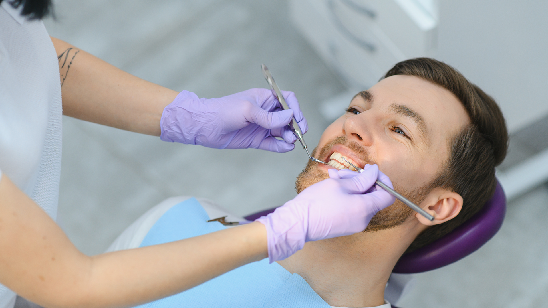
Diagnosis always begins with a clinical examination.
The dentist examines the teeth, assesses the accessible surfaces, the condition of the gums, oral hygiene, existing fillings and other factors that may affect the risk of tooth decay.
However, when posterior teeth are very close together, direct observation has its limitations.
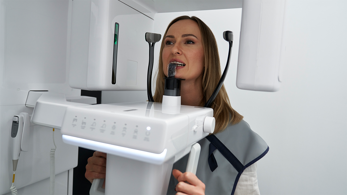
This is precisely where bitewing X-rays can be particularly useful. These images allow us to examine the crowns of the upper and lower teeth and, above all, the contact areas that are overlapped or obscured during a conventional visual examination.
On X-rays, a carious lesion may appear as an area of lower density in the enamel or dentine.
This does not mean, however, that everyone should have X-rays at every appointment. The need for X-rays and the frequency with which they are taken depend on individual risk, age, history of tooth decay, clinical findings and other circumstances.
There are also transillumination techniques that can serve as complementary methods in certain situations. No single test, on its own, can replace clinical judgement.
Does discovering a cavity always mean having a filling?
No.
This is an important distinction because modern dentistry does not treat all cases of tooth decay in the same way.
An initial lesion, where no cavitation has yet occurred, is not the same as a tooth surface where there is already significant physical loss of structure.
When the surface remains intact, certain lesions can be managed using non-restorative or minimally invasive treatments. The aim is to halt the disease process and prevent the lesion from progressing.
This may involve controlling biofilm, ensuring adequate fluoride exposure, reducing the frequency of sugar intake and clinical monitoring. In selected cases, there are also techniques such as resin infiltration for certain non-cavitated proximal lesions.
If cavitation is present or the condition has progressed to the point where a conservative approach is no longer feasible, a restoration may be necessary.
The depth observed on an X-ray is important, but it is not the only factor in the decision. That person’s risk of caries, the activity of the lesion, the feasibility of cleaning and the clinical characteristics of the surface also play a part.
Therefore, finding an image consistent with tooth decay does not automatically mean ‘drilling the tooth’.
So, does flossing prevent these cavities?
The question is a little more complex than simply answering ‘yes’ or ‘no’.
A toothbrush cleans various surfaces very well, but it does not actually reach the contact point between two teeth. Interdental hygiene helps to remove biofilm from areas that are difficult for a toothbrush to reach.
Depending on the space available, dental floss or another suitable interdental device may be used.
But preventing tooth decay does not depend on a single action.
The regular use of fluoride toothpaste, the quality of oral hygiene, the frequency with which foods and drinks high in fermentable sugars are consumed, salivary flow and individual risk are all part of the same issue.
A person may brush their teeth several times a day and still have factors that contribute to new lesions. Similarly, flossing does not make up for very frequent exposure to sugar.
The aim is to manage the process as a whole.
The aim is to manage the process as a whole.
It is useful to check your teeth regularly. This allows you to spot any changes in your gums, cracks, stains or obvious changes to the surface of your teeth.
However, it is no substitute for a clinical assessment.
Cavities between teeth are a good example of the difference between what we can see and what is actually happening in the mouth. They can exist without a visible cavity, without pain and without any changes that are obvious at home.
That is also why follow-up appointments are important, even when everything seems to be fine.
At Clinicalvor, the assessment combines a clinical examination with the diagnostic methods that each individual case actually requires. When there is an interproximal lesion, the aim is not simply to identify it. It is to determine how far it has progressed, whether it is active, and what the most conservative approach is to manage the problem.
Because, in the case of a cavity detected at an early stage, the most important question may not be how to restore the tooth, but how to avoid the need to do so.